 COA COA |
 MSDS MSDS |
 HPLC HPLC |
 NMR NMR |
| CAS No: | 601514-19-6 |
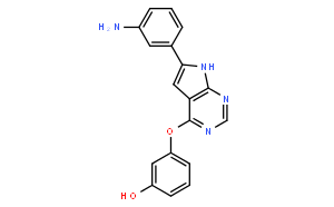
| Molecular formula(MF) | C18H14N4O2 |
| Molecular Weight(MW): | 318.33 |
| Alias |
| In vitro | DMSO | 64 mg/mL (201.04 mM) |
|---|---|---|
| Water | <1 mg/mL | |
| Ethanol | <1 mg/mL | |
| In vivo | 1% DMSO+30% polyethylene glycol+1% Tween 80 | 30 mg/mL |
| Description | TWS119 is a GSK-3β inhibitor with IC50 of 30 nM in a cell-free assay; capable of inducing neuronal differentiation and may be useful to stem cell biology. | ||||||||||||||||
|---|---|---|---|---|---|---|---|---|---|---|---|---|---|---|---|---|---|
| Targets |
|
||||||||||||||||
| In vitro |
Treatment of a monolayer of P19 cells with 1 μM TWS119 causes 30–40% cells to differentiate specifically into neuronal lineages based on counting of TuJ1 positive cells with correct neuronal morphology (up to 60% neuronal differentiation occurred through the standard EB formation protocol with concomitant TWS119 treatment). TWS119 tightly binds to GSK-3β (K D = 126 nM) which is quantified by surface plasmon resonance (SPR) and further demonstrates an IC50 of 30 nM. [1] TWS119 is found to potently induces neuronal differentiation in both mouse embryonal carcinoma and ES cells. [2] TWS119 treatment towards hepatic stellate cells (HSC) leads to reduced b-catenin phosphorylation, induces nuclear translocation of b-catenin, elevates glutamine synthetase production, impedes synthesis of smooth muscle actin and Wnt5a, but promotes the expression of glial fibrillary acidic protein, Wnt10b, and paired-like homeodomain transcription factor 2c. [3] TWS119 triggers a rapid accumulation of β-catenin (mean 6.8 -fold increase by densitometry), augments nuclear protein interaction with oligonucleotide containing the DNA sequences to which Tcf and Lef bind and sharply up-regulates the expression of Tcf7, Lef1 and other Wnt target genes including Jun, Ezd7 (encoding Frizzled-7), Nlk (encoding Nemo-like kinase). TWS119 induces a dose-dependent decrease in T cell-specific killing and IFN-g release associated with the preservation of the ability to produce IL-2. [4] A recent study indicates Wnt signaling is induced in polyclonally activated human T cells by treatment with TWS119. These T cells preserve a native CD45RA(+)CD62L(+) phenotype compared with control-activated T cells that progresses to a CD45RO(+)CD62L(-) effector phenotype and this occurs in a TWS119 dose-dependent manner. TWS119-induced Wnt signaling reduces T cell expansion as a result of a block in cell division, and impairs acquisition of T cell effector function as measured by degranulation and IFN-γ production in response to T cell activation. The block in T cell division may be attributed to reduced IL-2Rα expression in TWS119-treated T cells that lowers their capacity to use autocrine IL-2 for expansion. [5] |
||||||||||||||||
| Cell Data |
|
||||||||||||||||
| In vivo | A cell population that expressed low levels of CD44 and high levels of CD62L on the cell surface when 30 mg/kg of TWS119 is administered. [4] |